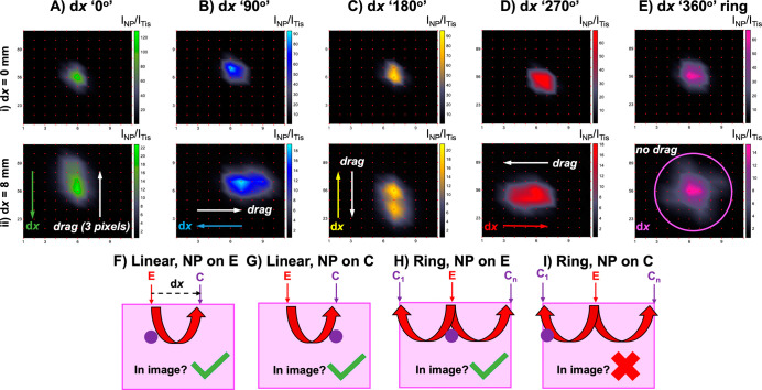
Figure 4.
Ring-collection ratiometric SESORS imaging as a means of removing linear offset-induced image drag. (A) Ratiometric images were collected of the PPY powder buried behind 3 mm of tissue across a 25 × 25 mm grid in 2.5 mm step sizes with linear offset magnitudes of (i) 0 and (ii) 8 mm. This was repeated three times with the sample being rotated through 360° in 90° steps to mimic rotation of the linear offset vector, and the resulting vectors were labeled with respect to the orientation of the vector in the initial set of images (dx “0°”) as (B) dx “90°”, (C) dx “180°”, and (D) dx “270°”. (E) Ratiometric images averaged across the four linear offset vectors to mimic a ring-collection offset at (i) 0 and (ii) 8 mm. (F–I) Schematics showing the effect of the optical geometry on the appearance of a through tissue SORS/SESORS image. Spectra were collected for 1 s using a laser power of 400 mW.

