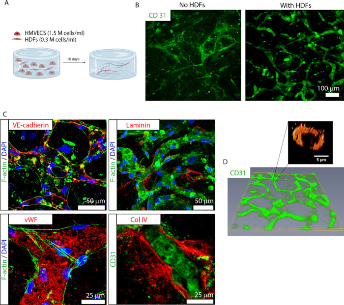
Figure 2.
Formation and characterization of a 3D vascular channel network composed of HMVECs and HDFs. (A) Illustration of the biofabrication process. Each 3D culture is composed of 300 μL of human fibrin containing HMVECs, at 1.5 M cells/mL and HDFs, at 0.3 M cells/mL, forming a ratio of 5:1. The culture is maintained for at least 10 DIV. Illustration made with biorender (https://biorender.com/). (B) Presence of HDFs within the culture is essential for the formation and maintenance of well-defined vessels. The micrograph on the left-hand side shows poor vessel formation in the absence of HDFs. In comparison, the presence of HDFs promotes the development of well-defined and interconnected vascular channels (right micrograph). The vascular channel formation is denoted by CD31 immunostaining (green). Scale bar represents 100 μm. (C) Characterization of the vessels’ phenotype with the traditional vascular markers (red in each respective panel): VE-cadherin (top left), laminin (top right), von Willebrand factor (vWF; bottom left), collagen type IV (col IV; bottom right), and CD31 (green, bottom right). F-actin for cytoskeleton (green) and 4′,6-diamidino-2-phenylindole (DAPI) for nuclei (blue) (top row and bottom left) are also shown. The scale bars represent 50 μm (top row) and 25 μm (bottom row). (D) Reconstruction of a micrograph depicting a 3D, interconnected vascular network, evidenced by CD31 immunostaining (green). The vascular network shows the presence of lumens with an average diameter of 5.9 μm.

