Figure 7.
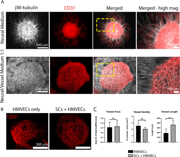
Vascular tissue development in the combined NV platform under different conditions. (A) Comparison of the neural and vascular tissue formation on platforms composed of SC-seeded scaffolds and hMVEC-laden fibrin cultured on neural medium (NM, top row) or 1:1 NM/VM (bottom row). NM promoted neurite outgrowth but was not sufficient to induce vessel formation, whereas 1:1 medium promoted simultaneous neuron proliferation, neurite outgrowth, and vessel formation, which was mostly circumscribed to the top of the neurosphere. Neurospheres labeled with βIII-tubulin (white) and vessels by CD31 (red). The images in the far right column show a high-magnification view of the yellow-dashed box marked in the merged column. Scale bars in the far left images represent 500 μm and apply to the two images to their respective right; scale bars in the far right images represent 100 μm. (B) Vessel formation (CD31, red) was similar on laminin-coated scaffolds (left) or SC-seeded scaffolds (right). Scale bars represent 500 μm. (C) Quantification of the vascular network on platforms with (gray bars) and without (black bars) SCs using the parameters: vessel area (left graph), vessel density (middle graph), and vessel length (right graph). The bar graphs represent mean ± SD from at least five replicates per condition in two independent experiments. Statistics were performed using an unpaired t-test, where ****p < 0.0001 and ns denotes not significant.

