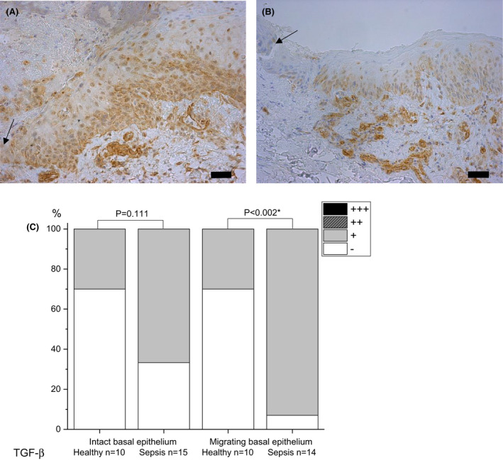
Fig. 3.

Immunohistochemical staining of transforming growth factor beta (TGF‐β) in skin on day 5 post wounding. Samples of septic patients (A) and healthy controls (B) are presented. The basal layer of migrating wound epithelium is stained more strongly in patient than control samples. Staining intensity is reported as absent (‐), mild (+), moderate (++) or strong (+++), and the percentages are shown (C). The arrow indicates the wound. The bar at the bottom of the figure is equal to 100 µm. Statistical significances between study groups are indicated by brackets and significant p‐values (p < 0.05) are marked with an asterisk.
