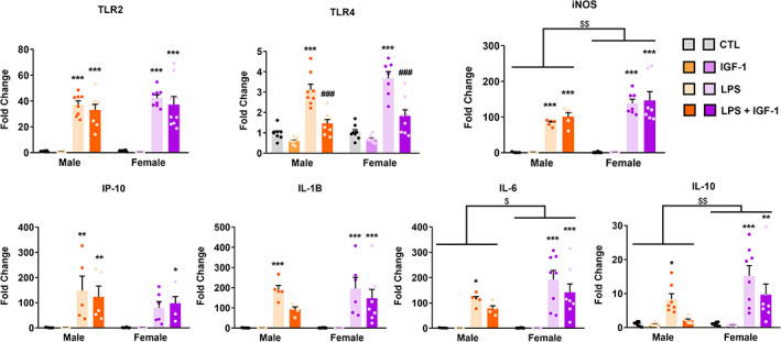
FIGURE 2.

Effect of IGF‐1 on the LPS‐induced inflammatory response in primary astrocytes from male and female mouse cortex. Astrocyte cultures were incubated for 24 h with 1 μg/ml LPS and/or 100 nM IGF‐1. The mRNA levels of TLR4, TLR2, iNOS, IP‐10, IL‐1β, IL‐6, and IL‐10 were measured on cell lysates by qRT‐PCR. The graphs show mRNA levels in cultures treated with vehicles (control), LPS, or LPS plus IGF‐1. Data represent the mean ± SEM. *p < .05; **p < .01; ***p < .001 effect of treatment in male and female astrocytes versus control groups and ### p < .001 versus LPS treated groups, measured by 2‐way ANOVA followed by Tukey's multiple comparisons post hoc test. $ p < .05; $$ p < .01 sex differences
