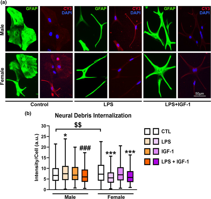
FIGURE 3.

IGF‐1 modulates phagocytosis of reactive male astrocytes. (a) Representative images of male and female astrocyte cultures treated for 24 h with vehicle, LPS and IGF‐1and then incubated for 1 h with Cy3 conjugated brain‐derived cellular debris (red). Cells were immunostained for GFAP (green) and cell nuclei were stained with DAPI. (b) Cy3 fluorescence intensity per cell in male and female astrocytes. Data are presented as median ± ranges. Significant differences *p < .05; ***p < .001, effect of treatment versus the control group of the same sex and ### p < .001 versus LPS treated groups, measured by the Kruskal‐Wallis test, followed by Dunn's multiple comparisons post hoc test, $$$ p < .001 basal sex differences measured by Mann–Whitney U test. N = 4–8 independent cultures
