Fig. 5.
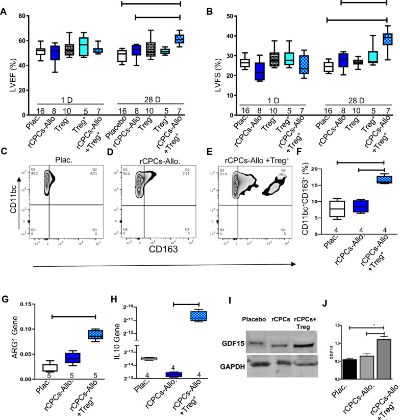
Tregs are essential for myocardial recovery after MI. Cardiac functional parameters were measured after intramyocardial injection of 1 million allogeneic rCPCs, Treg−, Treg+, rCPCs + Treg+ and placebo separately in a nude rat MI model. A and B represent LV ejection fraction and fractional shortening, respectively, at day 1 and day 28. Similarly, single-cell suspensions of total heart lysates were used for flow cytometric analysis of M2 elevation 5 days after intramyocardial injection of 1 million rCPCs, rCPCs + Treg+, or placebo. Representative images (C–E) and quantification (F). Left ventriculat tissue from the heart was used for RNA isolation and quantitative RT-PCR was performed to analyze the expression of G ARG-1, and H IL-10, respectively, from rCPCs, rCPCs + Treg+, and placebo-treated hearts. Numerical data are summarized as box and whisker plots with a median value (black bar inside box), 25th and 75th percentiles (bottom and top of box, respectively), and minimum and maximum values (bottom and top whisker, respectively). The number (n) of rats in each group indicated near the (up/below/on) each respective box and whisker plot. F and G Immunoblot analysis with quantification by densitometry revealed increased protein expression of GDF15 in nude rat myocardium after exposure to rCPCs + Treg + cells treatment relative to placebo and rCPCs (fold change relative to GAPDH listed below bands) (I, J)

