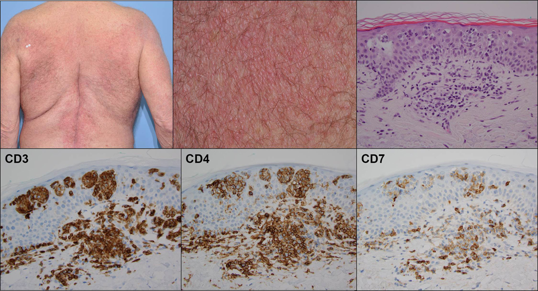
Figure 1:
Clinical photographs (low and high power) demonstrating broad symmetrical erythematous plaques with fine scale in a “bathing trunk” distribution (upper row left and middle). H&E stained histologic section demonstrates aggregates of atypical epidermotropic lymphocytes with superficial dermal perivascular involvement (upper row right). Immunohistochemical stains demonstrating staining of the infiltrate for CD3 and CD4, including many epidermotropic cells, and loss of staining for CD7 (lower row).

