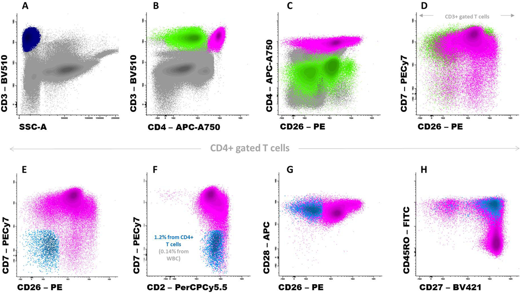
Figure 3:
A representative peripheral blood sample from an adult healthy donor stained with a combination of monoclonal antibodies designed for the identification of Sézary cells. Panel A shows total T cells (dark blue dots) while Panels B and C show the CD4+ T-cell (pink dots) and CD8+ T-cell subsets (green dots); non-T-cell events are displayed in gray. In Panel D, only T cells are displayed. Panels E-H show that a minor population of CD4+ T cells (displayed in light blue) has overlapping phenotypic features with Sézary cells: CD7-, CD26-, CD28+ and a mostly central memory (CD27+ and CD45RO+) phenotypic profile. Cells were stained using BulkLysis-based EuroFlow SOPs (www.euroflow.org); a total of 7×10⁶ leukocyte events are shown in Panels A-C.

