


Figure 13.
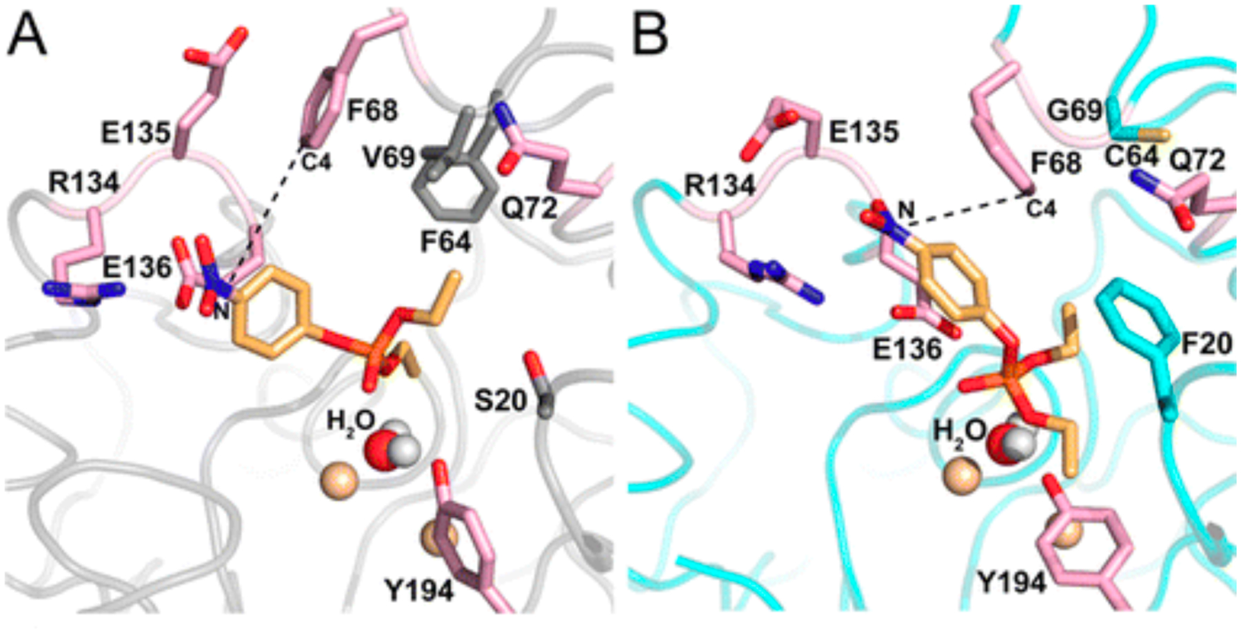
Paraoxon docked into the active site of wild-type AiiA (left) and a mutant enzyme with six mutations, three of which (S20F, V69G and F64C) reshape the active site. The position of the nucleophilic water and the two metal ions to which it is coordinated (gold spheres) are unchanged. In the wild-type enzyme, the substrate is not positioned correctly for in-line attack or water. Repositioning of Phe68 orients the substrate more appropriately. Reprinted with permission from Biochemistry 55(32):4583–93, 2106. Copyright 2016 American Chemical Society.