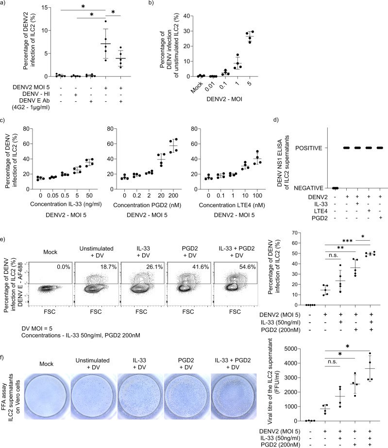
Fig. 2. Activated ILC2 are more permissive to infection and secrete infectious viral particles.
a ILC2 were exposed to live and heat inactivated DENV2 virus at a MOI of 5 for 2 h. In the blocking condition, dengue envelope protein (clone – 4G2 - 1 µg/ml) was mixed with dengue virus (MOI 5) and incubated for 1 h and was used to infect ILC2. Then, unbound virus was removed by washing cells twice and the cells were plated in fresh ILC2 media containing IL-2 for 48 h. Thereafter, intracellular DENV envelope (E) protein (Clone – 4G2) was checked by flow cytometry. (n = 4, Statistical significance was tested using one-way ANOVA with Tukey’s multiple comparison test, data representative of 3 independent experiments. P * < 0.05. b Unstimulated ILC2 and activated ILC2 (activated with PGD2 for 24 h) were exposed to live DENV2 virus at serial MOI from 0, 0.01, 0.1, 1 and 5 for 2 h. Then, unbound virus was removed by washing cells twice and the cells were plated in fresh ILC2 media containing IL-2 for 48 h. Intracellular DENV E protein was analysed by flow cytometry. (n = 4, data representative of two independent experiments). c ILC2 were activated through serial concentrations of IL-33 (0–50 ng/ml), PGD2 (0–200 nM) and LTE4 (0–100 nM) for 24 h. Then cells were incubated with dengue virus (MOI 5) for 2 h. Unbound virus was removed and the cells were plated in fresh ILC2 media containing IL-2 for 48 h. Thereafter, intracellular DENV E protein was analysed by flow cytometry. Data representative of three independent experiments. Error bars represent mean ± SD (n = 4). d DENV NS1 ELISA was performed of the supernatants of mock ILC2, infected unstimulated ILC2 and infected activated ILC2 (with IL-33 (50 ng/ml), PGD2 (200 nM) and LTE4 (100 nM) for 24 h). Data representative of two independent experiments (n = 5). e Activation of ILC2 with fixed concentrations of PGD2 (200 nM), IL-33 (50 ng/ml), LTE4 (100 nM) was performed for 24 h, followed by infection with dengue virus and then incubated for 48 h. Intracellular DENV E protein was analysed by flow cytometry. (n = 5, one-way ANOVA with Tukey’s multiple comparison test, data representative of three independent experiments. P * < 0.05, ** <0.01, *** <0.001, n.s. not significant.) f Supernatants of infected ILC2 of unstimulated and activated conditions (activated with IL-33 (50 ng/ml) and/or PGD2 (200 nM)) infected ILC2 (with MOI 5) were added on Vero cells and a Foci forming assay (FFA) was performed. Each focus is representative of an infected Vero cell. (n = 4, Statistical significance was tested using one-way ANOVA with Tukey’s multiple comparison test, data representative of 3 independent experiments. P * < 0.05, n.s. not significant). All error bars represent mean ± SD.

