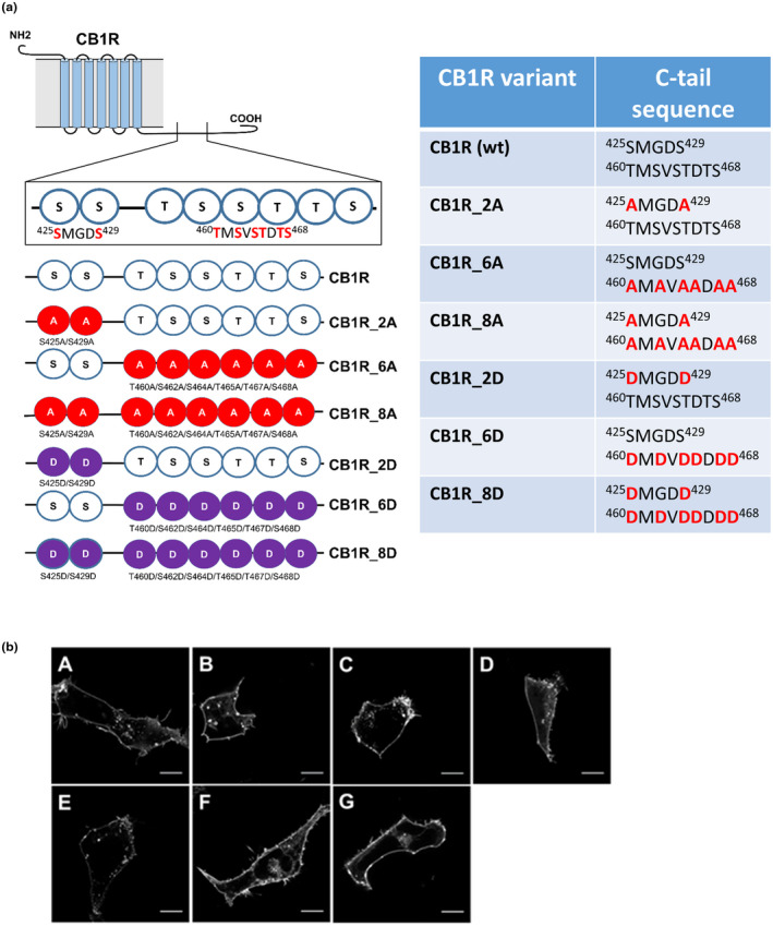
FIGURE 2.

Schematic depiction of CB1R mutants within the C‐tail and characterization of their cellular distribution. (a) List of CB1R mutants and their corresponding sequences. Two regions of CB1R: 425SMGDS429 and 460TMSVSTDTS468 contain serine/threonine residues that are possibly phosphorylated during desensitization of CB1R. CB1R C‐tail phosphorylation mutants were created according to the scheme: A—mutation into alanine, D—mutation into aspartic acid. (b) CB1R and mutant CB1Rs are predominantly localized on the cellular membrane. HEK293 cells were transiently transfected with CB1R‐YFP variant. Twenty‐four hours after transfection, cells were visualized using fluorescent microscope. A single confocal section through the equatorial plane of the cells is shown. Legend: (A) CB1R, (B) CB1R_2A, (C) CB1R_6A, (D) CB1R_8A, (E) CB1R_2D, (F) CB1R_6D, (G) CB1R_8D. Scale bar represents 10 μm
