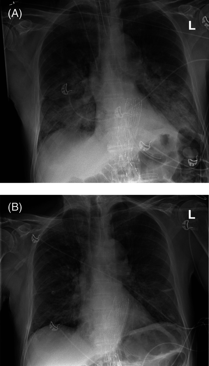
FIGURE 1.

Case 1 chest x‐ray. Panel A: 3 h:15 min after start of transfusion. Perihilar alveolar edema is present, with bilateral patchy consolidations visible. Panel B: 2‐days post‐transfusion: Improvement of consolidations in the lung fields bilaterally
