IMAGE 1.

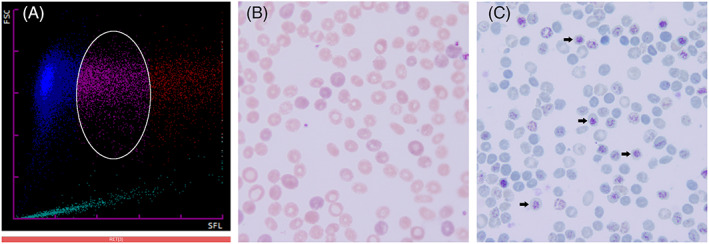
Morphologic and flow cytometry features of old reticulocytes. (A), Scattergram (Sysmex XN9000) (FSC, forward‐scattered light; SFL, side‐fluorescence light) demonstrating a population of low‐size reticulocytes with dim fluorescence (white circle). (B), Peripheral blood smear showing anisopoikilocytosis, polychromasia, and anisochromia (hematoxylin and eosin stain, original magnification ×1000). (C), Peripheral blood smear with arrows showing reticulocytes with dim fluorescence (low RNA content) on cresyl blue stain (original magnification ×1000)
