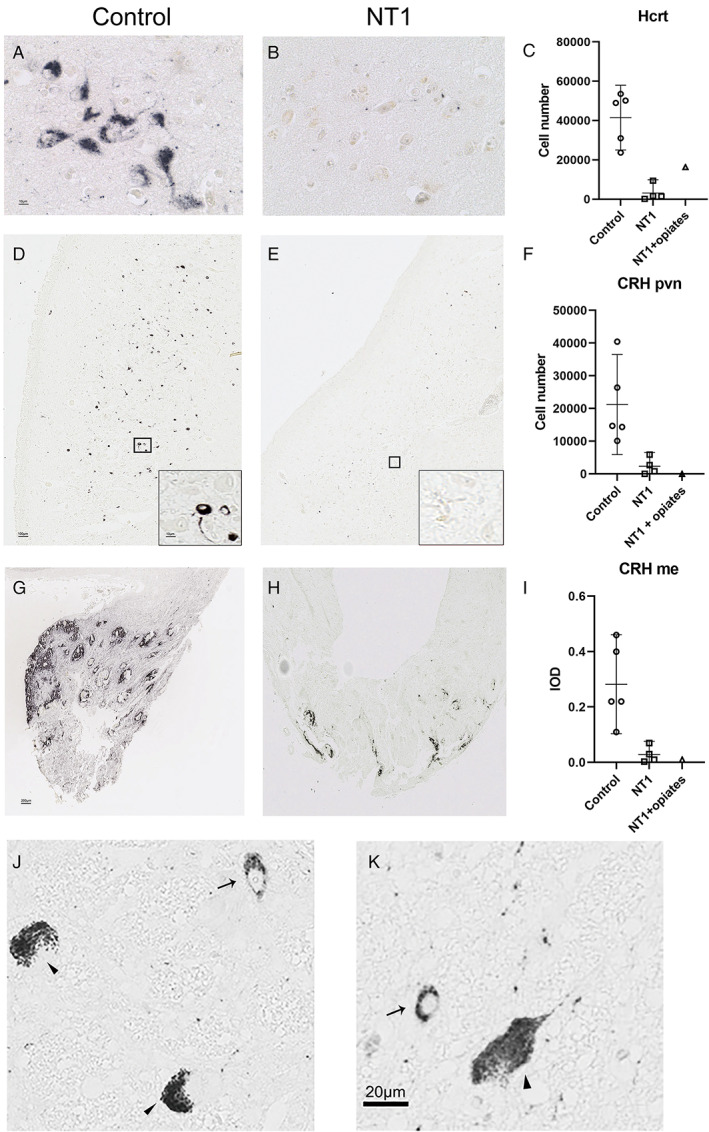
FIGURE 1.

Patients with narcolepsy type 1 (NT1) have not only a loss of 97% of hypocretin/orexin (Hcrt) neurons but also an 88% reduction of corticotropin‐releasing hormone (CRH) expressing neurons in the paraventricular nucleus (pvn) and an 91% reduction of CRH‐positive fiber staining in the median eminence (me). (A) Control and (B) NT1 Hcrt immunoreactive cells. (C) The total number of Hcrt neurons is more than 97% reduced in patients with NT1 compared to controls. (D) Control and (E) NT1 CRH immunoreactive cells in the pvn. (F) The total number of CRH neurons in the PVN is 88% lower in patients with NT1 than in controls. The subject with NT1 with chronic opiates (NT1 + opiates) also showed few CRH neurons. (G) Control and (H) NT1 fiber immunoreactivity in the median eminence. (I) The total optical density of CRH in the peak level of median eminence is 91% lower in NT1 than in controls. (J, K) Photomicrographs showing pigmented locus coeruleus neurons (indicated with an arrow head) and (J) an example of the positive CRH staining (indicated with an arrow) in the locus coeruleus of control, (K) a positive CRH staining (indicated with an arrow) in the locus coeruleus of the subject with NT1. The 6 μm sections of the locus coeruleus area contained up to 20 CRH neurons, in both, the controls and the patients with NT1. Scale bar represents 10 μm for A; 100 μm for D and 10 μm for insert; 200 μm for G and H, and 20 μm for K. Bar plots show the mean and the lower Bound‐Upper Bound of the 95% confidence intervals in C, F, and I. [Color figure can be viewed at www.annalsofneurology.org]
