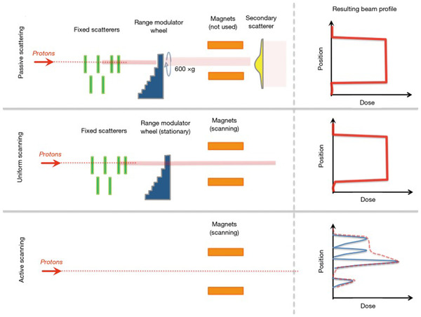
FIGURE 3.

An illustration of the hardware differences between passive scattering (top), uniform scanning (middle panel), and active scanning (a synonym for PBS) modalities. For active scanning (bottom row), the resulting radiation field is the sum of all of the individual spots, which may have different intensities. In the illustration, each spot contributes to the total dose indicated by the red dashed line. (Figure 2 from James SS, Grassberger C, Lu HM. Considerations when treating lung cancer with passive scattered or active scanning proton therapy. Transl. Lung Cancer Res. 2018;7(2):210–215)
