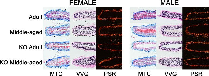
Figure 7.
Representative images of carotid artery histology. Formalin-fixed carotid cross sections were stained with Masson’s trichrome (MTC: blue, collagen; black, nuclei; red, smooth muscle), Verhoeff Van Gieson (VVG: black, elastin), and Picrosirius red (PSR: yellow/orange, thick collagen; green, thin collagen).

