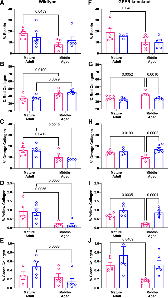
Figure 8.
Histological analysis of carotid cross sections by sex and age. Results from post hoc tests where P < 0.05 are indicated on each graph. n = 5 to 6 mice/group. In wild-type mice, elastin content (A) was decreased only in females. Picrosirius red staining revealed that red birefringent collagen (B) was similarly increased by age in both sexes, whereas orange (C) and yellow (D) collagen was decreased by aging in both sexes. Green (thin) collagen (E) was decreased only in males. In GPER knockout mice, elastin content (F) was reduced only in female mice. Red collagen (G) was increased with age only in females, whereas orange (H), yellow (I), and green (J) collagen was decreased with age only in females.

