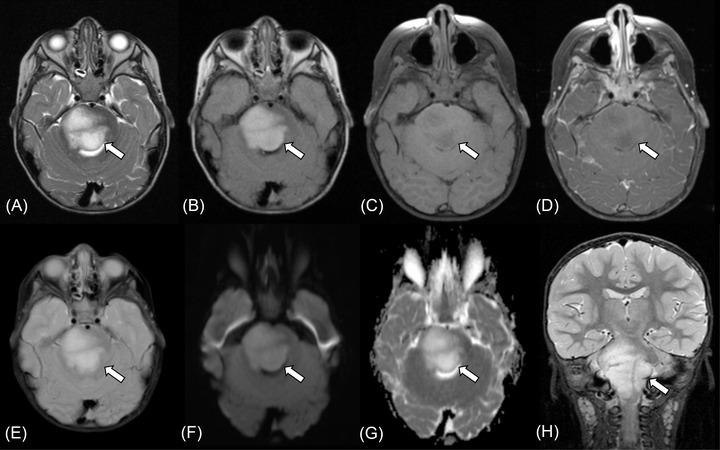
FIGURE 2.

Brainstem angiocentric glioma in a 2‐year‐old boy presenting with right facial weakness (case 1). MRI shows a 38 × 40 × 41 mm mass in the pontomedullary region (A‐H, arrows). The tumor shows high intensity on T2‐weighted image (A) and fluid‐attenuated inversion recovery image (B), low intensity on T1‐weighted image (C) without contrast enhancement (D). T2*‐weighted image does not show intratumoral calcification or hemorrhage (E). Diffusion restriction is not observed with the mean apparent diffusion coefficient value of 1.60 × 10⁻³ mm2/s (F, G). Fat‐suppressed coronal T2‐weighted image shows infiltrating growth of the tumor with an ill‐defined margin (H)
