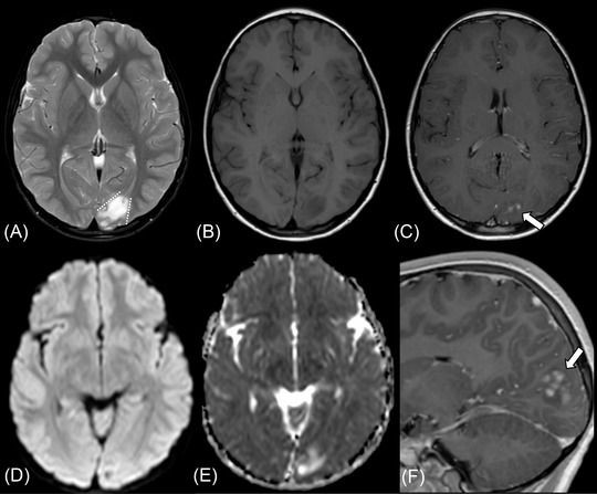
FIGURE 4.

Supratentorial angiocentric glioma in a 10‐year‐old male presenting with the first episode of epilepsy (case 3). The tumor shows high intensity on fat‐suppressed T2‐weighted image (A) and fluid‐attenuated inversion recovery image (not shown) and low intensity on T1‐weighted image (B). The stalk‐like sign is observed without evidence of atrophy in the surrounding brain parenchyma (A, dotted lines). Diffusion restriction is not observed with the mean apparent diffusion coefficient value of 1.34 × 10⁻³ mm2/s (D, E). Nodular enhancement is observed in the postcontrast sagittal T1‐weighted image (C, F, thick arrows)
