
FIGURE 1.
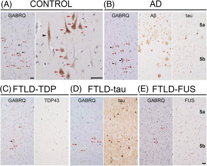
GABRQ immunopositive neurons in the ACC. GABRQ immunostaining is seen in VENs (black arrowhead) and surrounding pyramidal neurons (red arrow). Layer 5 GABRQ‐immunopositive neurons are seen in controls (A), whereas a reduced number of GABRQ‐positive neurons can be seen in donors with underlying TDP43 (C), tau (D) and FUS (E) pathology. In AD, a similar expression pattern of GABRQ‐positive neurons can be seen when compared to control (B). Adjacent images show pathology for each disease group. The same cases were used to denote typical GABRQ expression and pathology seen in each disease group (A, control case 11; B, AD case 3; C, FTLD‐TDP‐GRN case 6; D, FTLD‐tau‐MAPT case 9; E, FTLD‐FUS case 1). Scale bars represent 50 μm