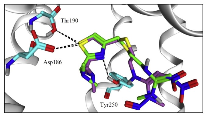
Fig. 3.
Binding mode of NZ, shown in green sticks and NZ-NO, shown in purple sticks and colored by element, into hH2R model. Amino acid residues are depicted in cyan colored sticks. Settled intermolecular interactions were shown in black dashed lines. Both ligands share a similar binding mode into the model’s pocket.

