
FIGURE 6.
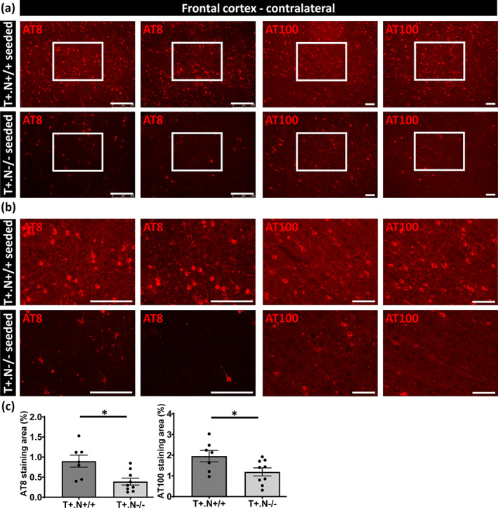
Decreased spreading of tau pathology in frontal cortex of tau‐seeded NLRP3‐deficient tau mice. (a) Representative images of AT8 (anti‐tau P‐S202/T205, left panels) and AT100 (anti‐tau P‐T212/S214, right panels) immunolabeling of the contralateral frontal cortex region of tau.NLRP3−/− (T+.N−/−) and tau.NLRP3+/+ (T+.N+/+), showing decreased levels of tau phosphorylation in the tau‐seeded T+.N−/− (lower panels) versus tau‐seeded T+.N+/+ (upper panels) mice. (b) Higher magnification of the contralateral frontal cortex region (corresponding to the white squared boxes in a) are shown. (c) Quantitative analysis showed significantly decreased AT8‐ and AT100‐positive area in the contralateral frontal cortex of the tau‐seeded T+.N−/− versus tau‐seeded T+.N+/+ mice. Data are shown as mean ± SEM (T+.N+/+: n = 7; T+.N−/−: n = 9; *p < .05; unpaired Welch's t‐test; AT8, bar = 250 μm; AT100, bar = 100 μm)