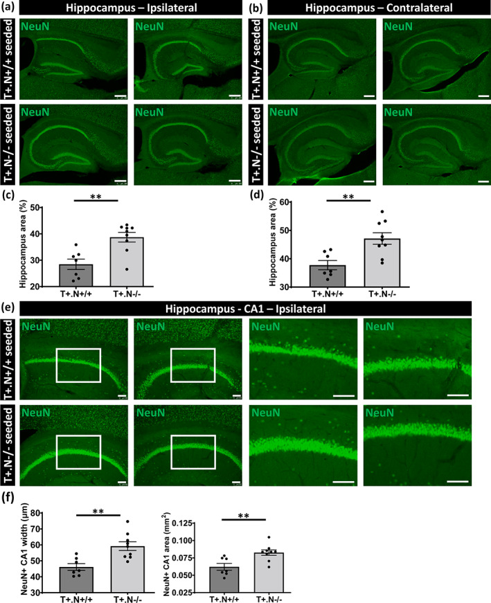
FIGURE 7.

Hippocampal atrophy and neurodegeneration in tau‐seeded NLRP3‐deficient tau mice. (a, b) Representative images of NeuN (neuronal nuclear antigen) staining of the ipsi‐ (a) and contra‐ (b) lateral hippocampus in well‐defined brain slices of exogenously seeded tau in tau.NLRP3+/+ (T+.N+/+, upper panels) and tau.NLRP3−/− (T+.N−/−, lower panels) mice, used for measuring the total hippocampal area. (c, d) Quantitative analysis showed significantly decreased total hippocampal area, in both ipsi‐ (c) and contra‐ (d) lateral sides, in tau‐seeded T+.N+/+ versus T+.N−/− mice. (e) Representative images of NeuN immunostaining of the ipsilateral hippocampal CA1 region of T+.N+/+ (upper panels) and T+.N−/− (lower panels) mice. Higher magnifications of selected areas (white squared boxes) are shown on the right. (f) Quantitative analysis showed significantly decreased width of the NeuN positive (NeuN+) neuronal layers in the CA1 region (left side) and decreased NeuN+CA1 area (right side) in the ipsilateral side in exogenously seeded tau T+.N+/+ versus T+.N−/− mice . Data are shown as mean ± SEM (T+.N+/+: n = 7; T+.N−/−: n = 9; **p < .01, ***p < .001; unpaired Welch's t‐test; hippocampus, bar = 250 μm; CA1 region, bar = 100 μm)
