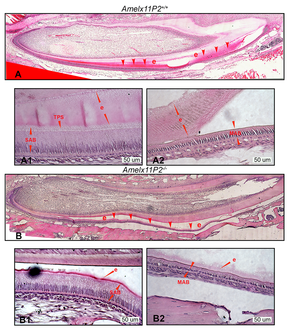
Fig.4.

Histological assessment of Amelx11P2−/− mouse ameloblasts and developing enamel matrix. A) H&E analysis shows that Amelx11P2+/+ enamel (e, indicated by red arrows) stained in pink grew thick through secretory stage of development and then disappeared at the end of maturation stage as a consequence of decalcification processing. A1) Enlarged image shows the properly differentiated secretory ameloblasts (SAB) with elongated cell body and “picket-fence” appearance Tomes’ processes (TPS) at the their apical pole, and organic enamel matrix (e) adhered to ameloblasts and dentin matrix. A2) Enlarged image shows the wild-type maturation stage ameloblasts (MAB) and the full thickness of mineralizing enamel matrix (e). B) Histological image shows that the enamel layer (e) in Amelx11P2−/− mouse incisor is thinner and prone to detach from dentin matrix. B1) In Amelx11P2−/− mouse incisor sagittal section, SAB is shorter and enamel layer (e) is thinner. B2) Both MAB and enamel matrix (e) are thinner as compared to that in Amelx11P2+/+ mouse incisor.
