Fig.8.

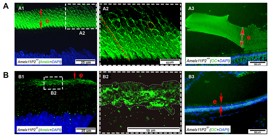
Assessment of the effects of 11P2 on amelogenin assembly in the developing enamel matrix. Super-resolution confocal microscopic image shows that amelogenin was detected in the secretory stage enamel matrix (e) (A1). Amelogenin positive signal either distributed along the long-axis of enamel rods like fibrils (indicated by the dotted red line) or encompassed the objects in the shape of hexagon (indicated by dotted red hexagon) in Amelx11P2+/+ enamel matrix (A2). A3) OC antibody immunostaining analysis shows the positive signal (in green) colocalized with the aligned enamel rods in enamel matrix (e). B1) There is less amelogenin immunostaining signal in the thin Amelx11P2−/− enamel matrix (e). B2) Enlarged image shows that amelogenin distributed in enamel matrix without a pattern. B3) No OC positive immunostaining signal was found in Amelx11P2−/− mouse enamel matrix (e).
