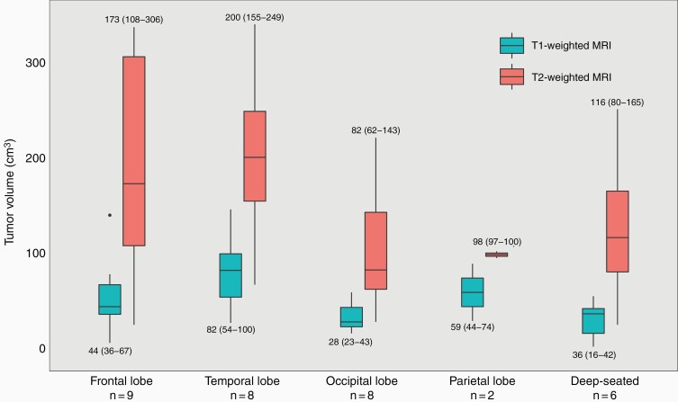
Figure 1.
Median [interquartile range (IQR)] contrast-enhancing tumor volume in T1-weighted magnetic resonance imaging (MRI) and median (IQR) volume of tumor-associated non-enhancing hyperintense lesions in T2-weighted and/or fluid-attenuated inversion recovery (FLAIR) MRI according to tumor location in 33 patients diagnosed with butterfly glioblastoma between 01/01/2007 and 31/12/2014.

