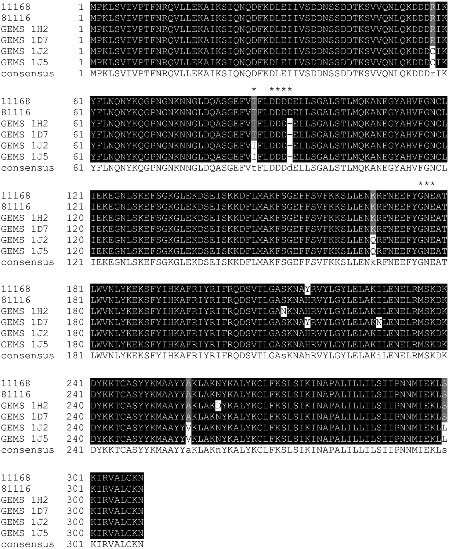
Figure 2. Multiple sequence alignment of Campylobacter GEMS PglI proteins.

The DXDD motif as found in the reference strains Cj-11168 and Cj-81116 (where glycosylation of the N-glycan takes place to form the full length heptasaccharide) as well as the 177Gly-Asn-Glu179 motif that corresponds to the x-Glu-Asp motif containing the catalytic base in other related GTases are highlighted by asterisks. Highlighted in black are amino acids that are invariable, gray highlighting indicates amino acid conservation in at least 50% of the sequences, white indicates a non-conserved amino acid, and (−) marks a deletion at this position.
