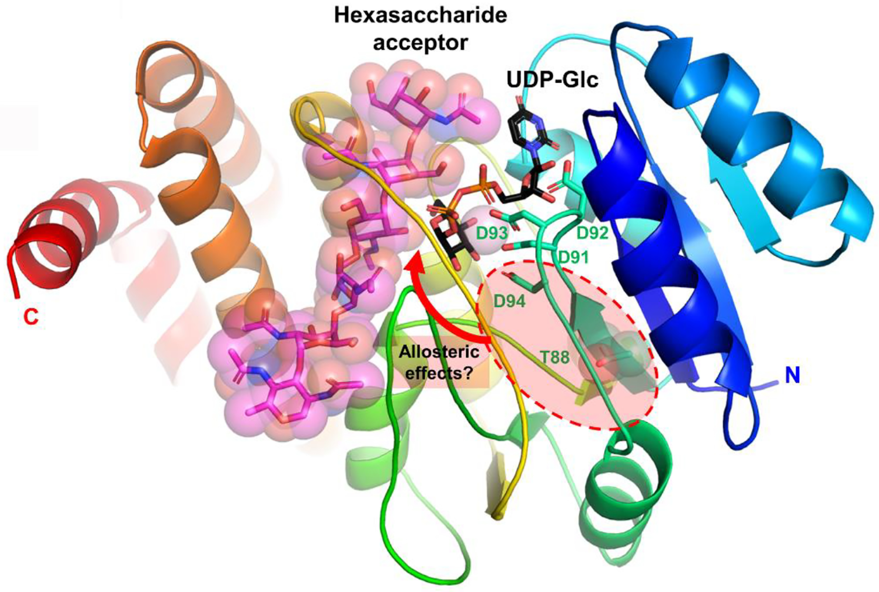
Figure 5. Structural model of PglI.

Cartoon representation of secondary structure elements with rainbow coloring starting with blue at the N-terminus and red at the C-terminus. UDP-Glc donor (black), hexasaccharide acceptor (magenta), the Asp91-X-Asp93 motif, Asp94 and Thr88 are all drawn in stick representation.
