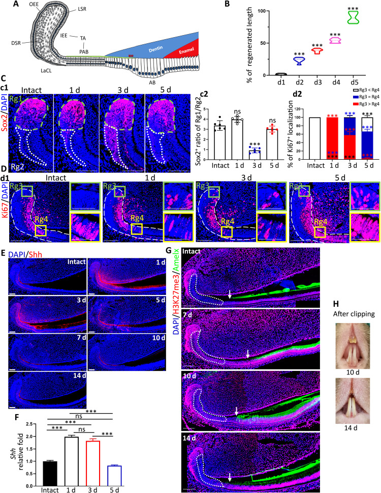
Fig. 1. Identification of injury-induced cell fate alterations and spatiotemporal expression of Shh and H3K27me3 status.
(A) Schematic illustration of dental epithelium of adult mandibular incisors. (B) Statistical data of the regeneration ratio of mandibular incisors at different time points after clipping at the gingival level. (C) c1: Immunofluorescent images of Sox2+ DESCs in LaCL of intact or defect incisors. c2: The ratio of Sox2+ DESCs in the upper layer of LaCL over that at the bottom of LaCL at different time points after injury. (D) d1: Immunofluorescent images of Ki67+ proliferative cells. d2: Statistical data of d1. (E) Representative IF images of Shh. The labels of days indicated the days after clipping. (F) Statistic RT-qPCR data of Shh in healthy and different stages after clipping. (G) Representative IF images of H3K27me3 and Amelx in intact condition and 7, 10, and 14 days after clipping. Dotted areas indicate the IEE region. The arrow indicates the deposit start site of Amelx. (H) Representative photographs of incisors at 7 and 10 days after clipping. All representative images and data (means ± SEM) were generated from at least six slices from five mice for each group. ns, no statistical significance; *P < 0.05, **P < 0.01, and ***P < 0.001. The dot-square contoured regions were shown in the right as high-magnification images. OEE, outer enamel epithelium; DSR, dense stellate reticular layer; LSR, loose stellate reticular layer; BM, basal membrane; AB, ameloblasts. Scale bar, 50 μm.

