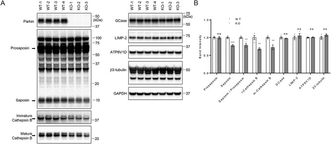
Figure 7.
Saposin and cathepsin B levels are reduced in Parkin knockout (KO) mouse brain; (A) 20 μg of tissue lysates isolated form whole brains from 12-week-old WT and Parkin KO mice were analyzed with immunoblot analysis using indicated antibodies. Representative immunoblot data are shown. (B) Quantification of immunoblots. Protein band intensities were normalized with GAPDH and compared with WT. Two-tailed unpaired t-test was used to compare KO to WT mice (n = 4 for WT; n = 3 for KO) (**P < 0.01; ***P < 0.001; ns, not significant).

