Figure 4.
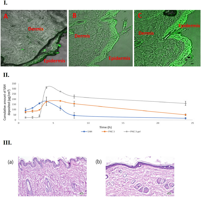
(I) Confocal laser scanning micrographs (CLSMs), obtained after 8 h following the application of Rhodamine B dye aqueous solution (A), Rhodamine B dye-loaded PNE 3 (B), and Rhodamine B dye-loaded PNE 3 gel (C), (II) The plot of the amount of SAH deposited per unit area in rat skin after the application of SAH, PNE 3, and PNE 3 gel, and (III) Histopathological photomicrographs of skin tolerance study; showing histopathological sections (hematoxylin- and eosin-stained) of (a) Untreated rat’s skin (group A); showing a normal histological structure of the epidermis and the skin appendages, and (b) Rat’s skin treated with SAH-loaded PNE 3 gel (group B) showing an apparent normal architecture of the epidermis and the dermis at 50 µm.

