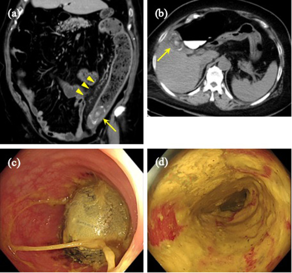
FIGURE 1.

Colonic gallstone ileus. (a) Computed tomography showing a 46 × 30 mm elliptical calcification (yellow arrow) lodged in the sigmoid‐descending colon junction, and dilation of the oral colon with pericolic inflammation (yellow arrowhead). (b) A gallstone of the same measurements was confirmed in a computed tomography scan 5 years prior (yellow arrow) (c) Colonoscopy showing the gallstone impacted at the sigmoid‐descending colon junction. (d) The mucosa proximal to the obstruction appears red and edematous, compatible with obstructive colitis
