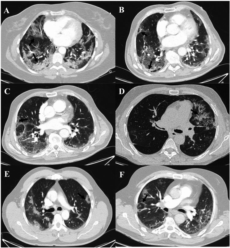
Figure 2. CT features of organising pneumonia in COVID-19 pneumonia patients.
(A) Bilateral subpleural and peribronchovascular consolidations (black arrow). (B) Subpleural linear consolidation (black arrow). (C) Parenchymal bands (black arrow). (D) Crazy paving pattern (ground-glass opacities with inter and intralobular septal thickening) (black arrow). (E) Perilobular opacities (ill-defined perilobular linear opacities, thicker than the thickened interlobular septa with an arch shape) (black arrow). (F) Reversed halo (attol) sign (central ground-glass opacity surrounded by denser consolidation of crescentic shape) (black arrow).

