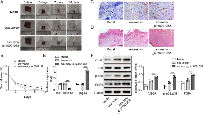
Fig. 5.
Mmu_circ_0001052 from ADSCs-derived exosomes promoted wound healing in DFU via miR-106a-5p and FGF4/p38MAPK pathway. A wound healing in DFU with exosomes treatment. B the broken line diagram of wound healing in DFU mice. C the vessel formation under Immunofluorescence. D HE staining. E the expression of miR-106a-5p and FGF4 in DFU mice. F, the level of FGF4/p38 pathway via western blot. All data were obtained from at least three replicate experiments. *P < 0.05, **P < 0.01. N = 5. One-way ANOVA was used to analyze the differences among groups. Afterward, post-pairwise comparison was conducted by Dunnett’s multiple comparison test. Comparison among multiple groups at different time points was analyzed by repeated measures ANOVA

