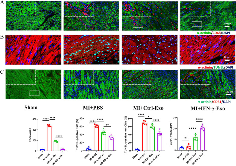
Fig. 4.
IFN-γ-Exos were better at inhibiting apoptosis or promoting angiogenesis than IFN-γ-Exo in vivo. (A) Representative fluorescence images of macrophages in the border zone of ischemic hearts stained with CD68 (red) and α-actinin (green) (3 random fields per anima). (B) Representative photographs showing the TUNEL-positive cells (green) in the heart tissue (red) among the different groups. Quantitative analysis of the apoptotic rate at the border zone in CMs and non-CMs among the different groups (3 random fields per animal). (C) Representative fluorescence images of blood vessels in the border zone of ischemic hearts stained with CD31 (red) and α-actinin (green) (3 random fields per animal). Data are presented as mean ± SEM. Statistical analysis was performed with one-way ANOVA followed by Bonferroni’s correction. *P < 0.05; **P < 0.01; ***P < 0.001; ****P < 0.0001

