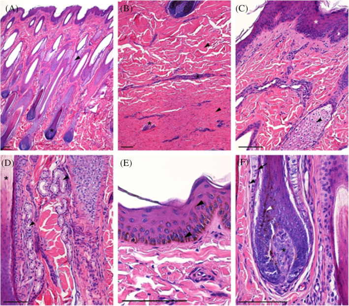
FIGURE 1.

Normal horse skin. (A) Papillary layer of the dermis with hair follicles (asterisk) and accompanying sebaceous glands (arrowhead); (B) Reticular layer of the dermis with thick bundles of collagen fibers (arrowheads) and blood vessels (asterisk); (C) Superficial part of the dermis with epidermis (asterisk) and hair sebaceous gland; (D) Deeper part of the papillary layer with sweat gland (arrowhead) between hair follicles (asterisk); (E) Epidermis (keratinized stratified squamous epithelium) with numerous melanin granules (arrowhead); (F) Hair bulb with pigmented cells (white arrowhead) attached to the dermal papilla (asterisk) and covered by hair follicle with epithelial hair root sheath (arrow) and dermal sheath (arrowhead); H&E staining, scale bar = 100 μm
