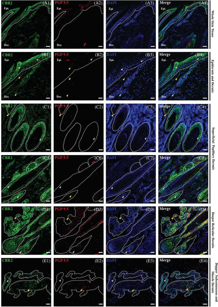
FIGURE 3.

Triple confocal microscopic in‐situ expression of CBR2 (green), PGP 9.5 (red) and DAPI (blue) in the whole‐skin tissue under 20x magnification; scale bars = 50 μm (A1‐A4). Below, a set of images presenting the layers of the equine skin, with emphasis on their respective epidermal and dermal compartments. Epidermis (Epi.: stratum basale, granulosum, spinosum and corneum; B1‐B2) and dermis (B1‐B2) and dermis (Der.: upper and lower superficial papillary dermis—C1‐C8, deeper reticular dermis—D1‐D4, and deep subcutaneous and muscle dermis—E1‐E4) with their respective compartments. The specific region of interest (white dotted lines) and randomly selected example regions with single CBR2 (gray arrows) and double positive in co‐localization with PGP 9.5 (yellow arrows) specific immunoreactivity are indicated. Images were taken under 40x magnification, scale bars = 20 μm
