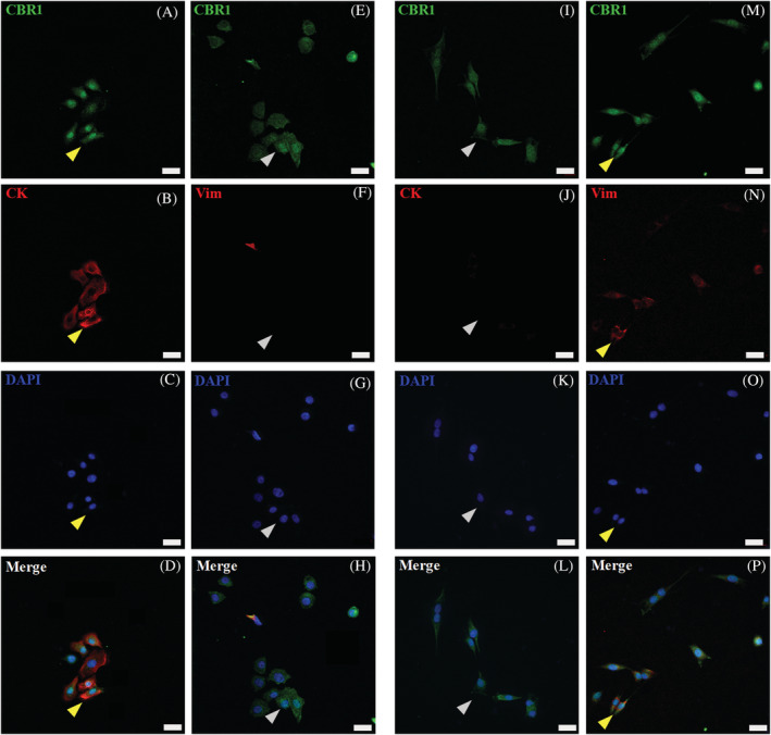
FIGURE 7.

CBR1 (CBR1‐green) confocal microscopic results, showing images of equine primary skin‐derived cultures of keratinocytes (A‐H) and fibroblasts (I‐P). CBR1 immunoreactivity in keratinocytes (A and E) and fibroblasts (I and M). For keratinocyte and fibroblast immunophenotyping, co‐labeling of CBR1 with specific epithelial pan‐cytokeratin (CK‐red) (B) and fibroblast vimentin (Vim‐red) (N) markers was performed (yellow arrows indicate double positive cells). Inverse staining of Vim‐red on keratinocytes (F) and CK‐red on fibroblast (J) with CBR1 was additionally performed to distinguish both primary cell types (gray arrows indicate single positive cells). The DAPI (blue) was used for cell nucleus counterstaining; images were taken under 40x magnification; scale bars = 20 μm
