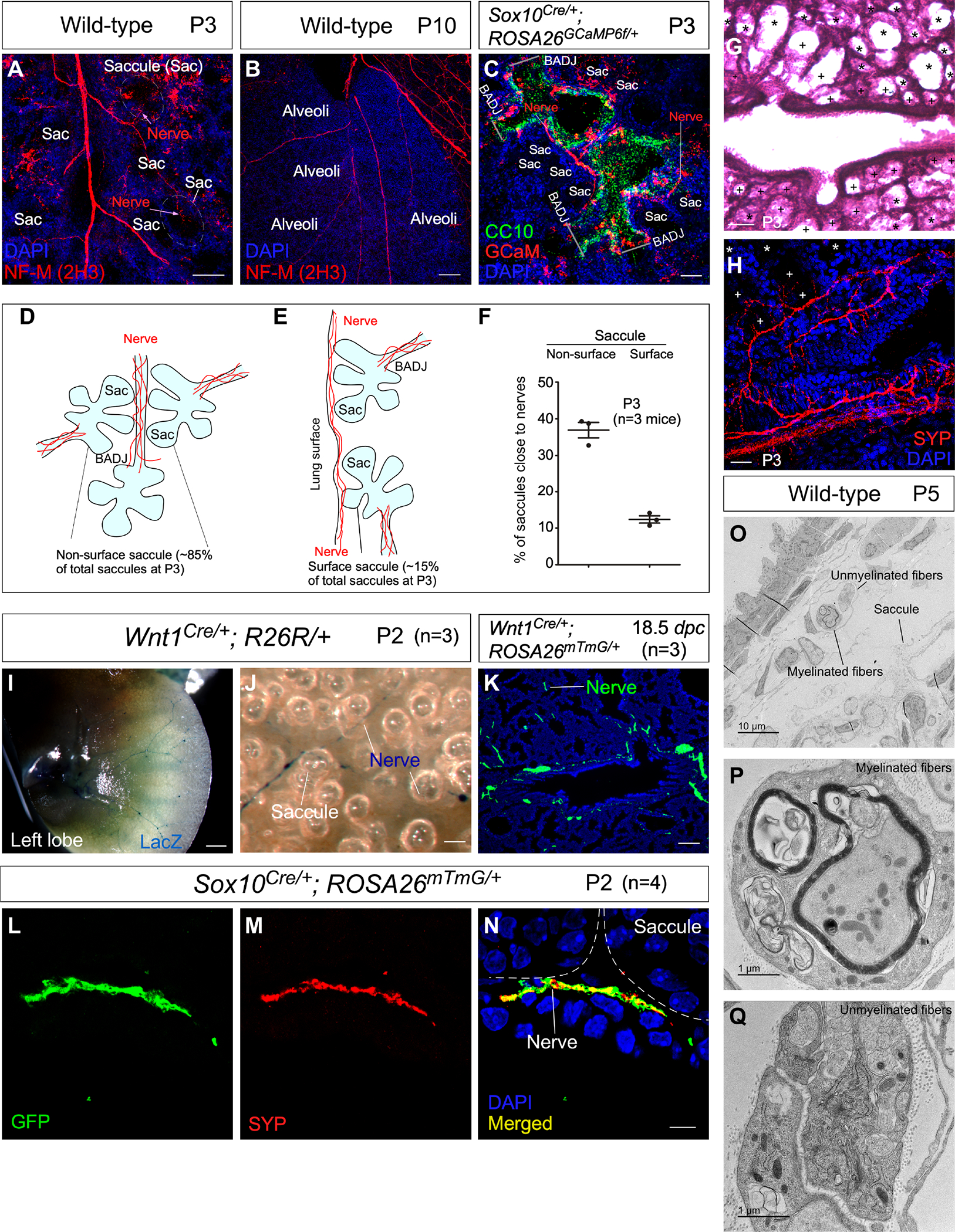
Figure 1. Mouse saccules are innervated by neural crest descendants.

(A-C) Immunofluorescence of mouse lung sections stained with anti-NF-M (2H3) to trace autonomic nerves at postnatal (P) day 3 and 10. Saccules (Sac) and alveoli adjacent to nerves were identified by their morphology. Autonomic nerves were also visualized by GCaM expression in Sox10Cre/+; ROSA26GCaMP6f/+ lungs at P3, in which GCaM was activated from ROSA26GCaMP6f in neural crest descendants (SOX10+) after their migration. The airway epithelium was marked by CC10 expression in club cells, aiding the identification of the bronchoalveolar duct junction (BADJ) that separated the airways and saccules. The addition of a clearing procedure facilitated visualization of nerve fibers.
(D, E) Schematic diagram of the passage of autonomic nerves to reach both surface and non-surface saccules.
(F) Quantification of the % of saccules that are adjacent to autonomic nerves of wild-type mouse lungs at P3. Saccules were divided into the surface and non-surface groups as shown in (D, E).
(G) Hematoxylin-and-eosin (H&E) staining of lung sections from control mice at P3. Saccules marked by (+) were counted as saccules close to nerves whereas saccules marked by (*) were counted as saccules not close to nerves.
(H) Immunofluorescence of lung sections from control mice stained with anti-SYP. Saccules marked by (+) were counted as saccules close to nerves whereas saccules marked by (*) were counted as saccules not close to nerves.
(I, J) Surface view of LacZ-stained left lobes of mouse lungs from Wnt1Cre/+; R26R/+ mice (n=3) at P2. LacZ expression was induced from R26R in neural crest descendants (WNT1+) prior to their migration.
(K) Immunofluorescence of lung sections from Wnt1Cre/+; ROSA26mTmG/+ mice (n=3) at 18.5 days post coitus (dpc) stained with anti-GFP. GFP expression was generated from ROSA26mTmG in WNT1+ cells. Nerve fibers (GFP+) were in close proximity to the developing saccules.
(L-N) Immunofluorescence of lung sections from Sox10Cre/+; ROSA26mTmG/+ mice (n=4) at P2 stained with anti-GFP and anti-SYP. GFP expression was produced from ROSA26mTmG in SOX10+ cells. Autonomic nerve fibers (GFP+SYP+) adjoined developing saccules.
(O-Q) Transmission electron microscopy (TEM) of wild-type lungs to visualize autonomic nerve fibers and their relationship to saccules.
Scale bars, 100 μm (A), 100 μm (B), 50 μm (C), 100 μm (G), 25 μm (H), 1 mm (I), 25 μm (J), 100 μm (K), 5 μm (L-N).
