
Figure 3. Autonomic nerve activity regulates alveolar formation.
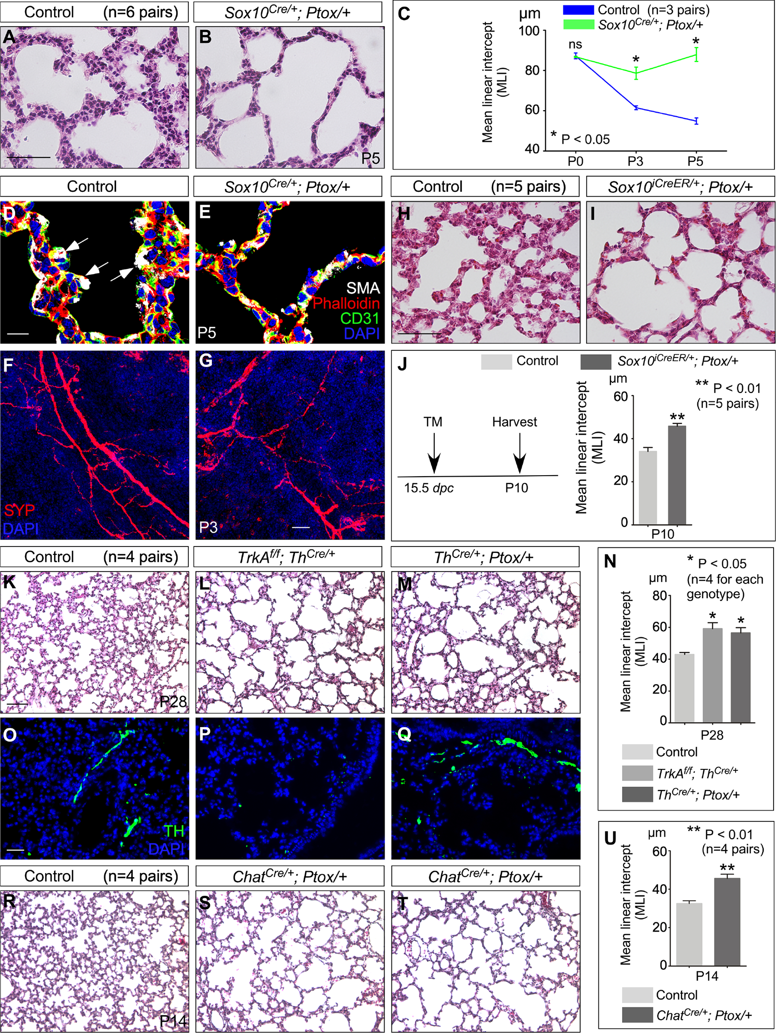
(A, B) H&E staining of lung sections from control and Sox10Cre/+; Ptox/+ mice (n=6 pairs) at P5.
(C) Measurement of the MLI in control and Sox10Cre/+; Ptox/+ lungs (n=3 pairs) at P0, P3 and P5.
(D, E) Immunofluorescence of lung sections from control and Sox10Cre/+; Ptox/+ mice (n=3 pairs) stained with anti-SMA (smooth muscle actin), anti-CD31 and phalloidin at P5. CD31 labels endothelial cells and phalloidin binds to F-actin. Arrows point to secondary septation.
(F, G) Immunohistochemical analysis of control and Sox10Cre/+; Ptox/+ mouse lungs at P3. Immunoreactivity of SYP, which marked nerve fibers, showed no apparent difference between control and mutant lungs.
(H, I) H&E staining of lung sections from control and Ptox-mutant mice (conditional induction by tamoxifen).
(J) Measurement of the MLI in control and Sox10iCreER/+; Ptox/+ lungs (n=5 pairs) at P10. Sox10iCreER/+; Ptox/+ mice were injected with tamoxifen at 15.5 dpc and lungs were collected at P10.
(K-M) H&E staining of lung sections from control, TrkAf/f; ThCre/+ and ThCre/+; Ptox/+ mice at P28.
(N) Measurement of the MLI in control, TrkAf/f; ThCre/+ and ThCre/+; Ptox/+ lungs at P28 (n=4 for each genotype).
(O-Q) Immunohistochemical analysis of control, TrkAf/f; ThCre/+ and ThCre/+; Ptox/+ mouse lungs at P28. Immunoreactivity of TH, which marked sympathetic nerves, was reduced in TrkAf/f; ThCre/+ lungs but not in ThCre/+; Ptox/+ lungs.
(R-T) H&E staining of lung sections from control and ChatCre/+; Ptox/+ mice at P14.
(U) Measurement of the MLI in control and ChatCre/+; Ptox/+ lungs at P14 (n=4 pairs).
Scale bars, 100 μm (A, B), 10 μm (D, E), 50 μm (F, G), 100 μm (H, I), 100 μm (K-M, R-T), 25 μm (O-Q). All values are mean ± SEM. (*) p<0.05; (**) p<0.01; ns, not significant (unpaired Student’s t-test and one-way ANOVA).