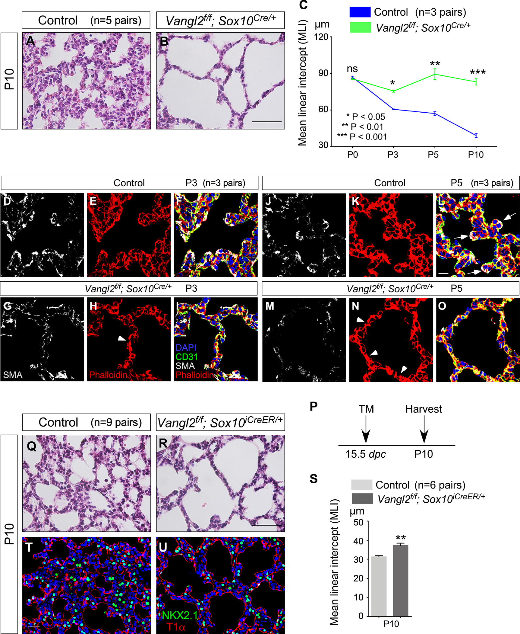
Figure 4. A signaling cascade of planar cell polarity controls alveolar formation.

(A, B) Hematoxylin-and-eosin (H&E) staining of lung sections from control and Vangl2f/f; Sox10Cre/+ mice (n=5 pairs) at postnatal (P) day 10.
(C) Measurement of the mean linear intercept (MLI) in control and Vangl2f/f; Sox10Cre/+ lungs (n=3 pairs) at P0, P3, P5 and P10. The early phenotypes suggest that the requisite expansion and migration of myofibroblasts prior to alveologenesis are disrupted in the absence of PCP signaling in autonomic nerves.
(D-O) Immunofluorescence of lung sections from control and Vangl2f/f; Sox10Cre/+ (n=3 pairs for each stage) mice stained with anti-SMA (smooth muscle actin), anti-CD31 and phalloidin at P3 and P5. CD31 labels endothelial cells and phalloidin binds to F-actin. Arrows point to sites of secondary septation. Arrowheads indicate disorganized cytoskeleton in the mutant lungs.
(P) Schematic diagram of the time course of tamoxifen administration and tissue harvest for control and Vangl2f/f; Sox10iCreER/+ mice.
(Q, R) H&E staining of lung sections from control and Vangl2f/f; Sox10iCreER/+ mice (n=9 pairs) at P10.
(S) Measurement of the MLI in control and Vangl2f/f; Sox10iCreER/+ lungs (n=6 pairs) at P10.
(T, U) Immunofluorescence of lung sections from control and Vangl2f/f; Sox10iCreER/+ mice (n=3 pairs) stained with anti-NKX2.1 and T1α at P10. NKX2.1 labeled epithelial cells while T1α marked alveolar type I cells.
Scale bars, 100 μm (A, B), 10 μm (D-O), 100 μm (Q, R), 25 μm (T, U). All values are mean ± SEM. (*) p<0.05; (**) p<0.01; ns, not significant (unpaired Student’s t-test).
