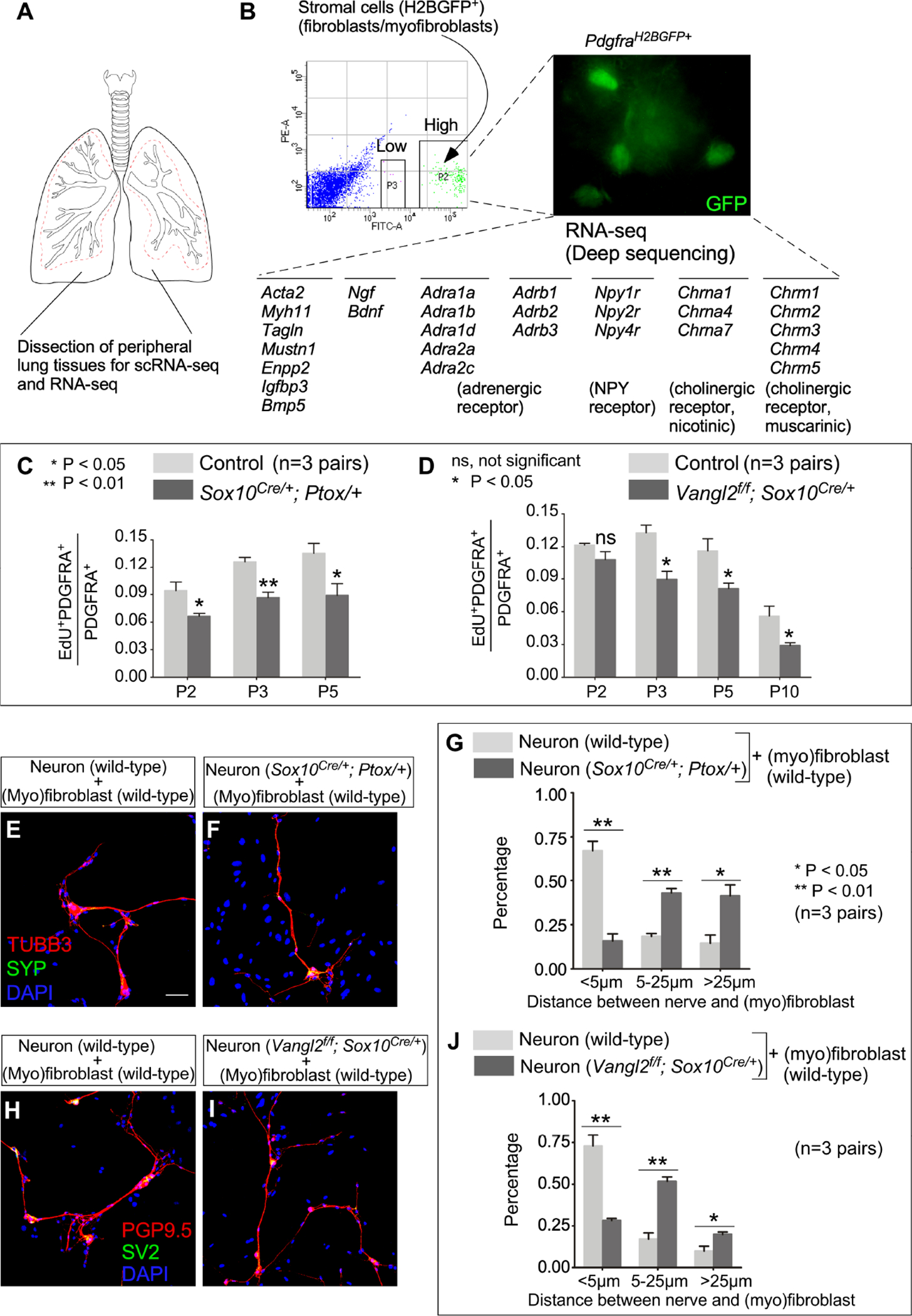
Figure 7. A functional interaction between alveolar fibroblasts/myofibroblasts and autonomic nerves is essential for alveologenesis.

(A) Schematic diagram of dissection of peripheral lung tissues for scRNA-seq and RNA-seq.
(B) RNA-seq analysis of sorted murine myofibroblasts. (Myo)fibroblasts (GFP+) were isolated from the lungs of PdgfraH2BGFP/+ mice at postnatal (P) day 3 by fluorescence-activated cell sorting (FACS). A partial list of genes involved in neurotrophin signaling and synaptic transmission in sympathetic and parasympathetic nerves were shown.
(C) Quantification of the ratio of proliferating (myo)fibroblasts (EdU+PDGFRA+) to (myo)fibroblasts (PDGFRA+) in control and Sox10Cre/+; Ptox/+ lungs (n=3 pairs) at P2, P3 and P5.
(D) Quantification of the ratio of proliferating (myo)fibroblasts (EdU+PDGFRA+) to (myo)fibroblasts (PDGFRA+) in control and Vangl2f/f; Sox10Cre/+ lungs (n=3 pairs) at P2, P3, P5 and P10.
(E, F) Immunofluorescence of cultured neurons and (myo)fibroblasts.
(G) Quantification of the distance between nerves and (myo)fibroblasts, which was measured as the shortest distance between the nuclei of (myo)fibroblasts and the nerve fibers.
(H, I) Immunofluorescence of cultured neurons and (myo)fibroblasts.
(J) Quantification of the distance between nerves and (myo)fibroblasts, which was measured as the shortest distance between the nuclei of (myo)fibroblasts and the nerve fibers.
Scale bars, 100 μm (E, F, H, I). All values are mean ± SEM. (*) p<0.05; (**) p<0.01; ns, not significant (unpaired Student’s t-test).
