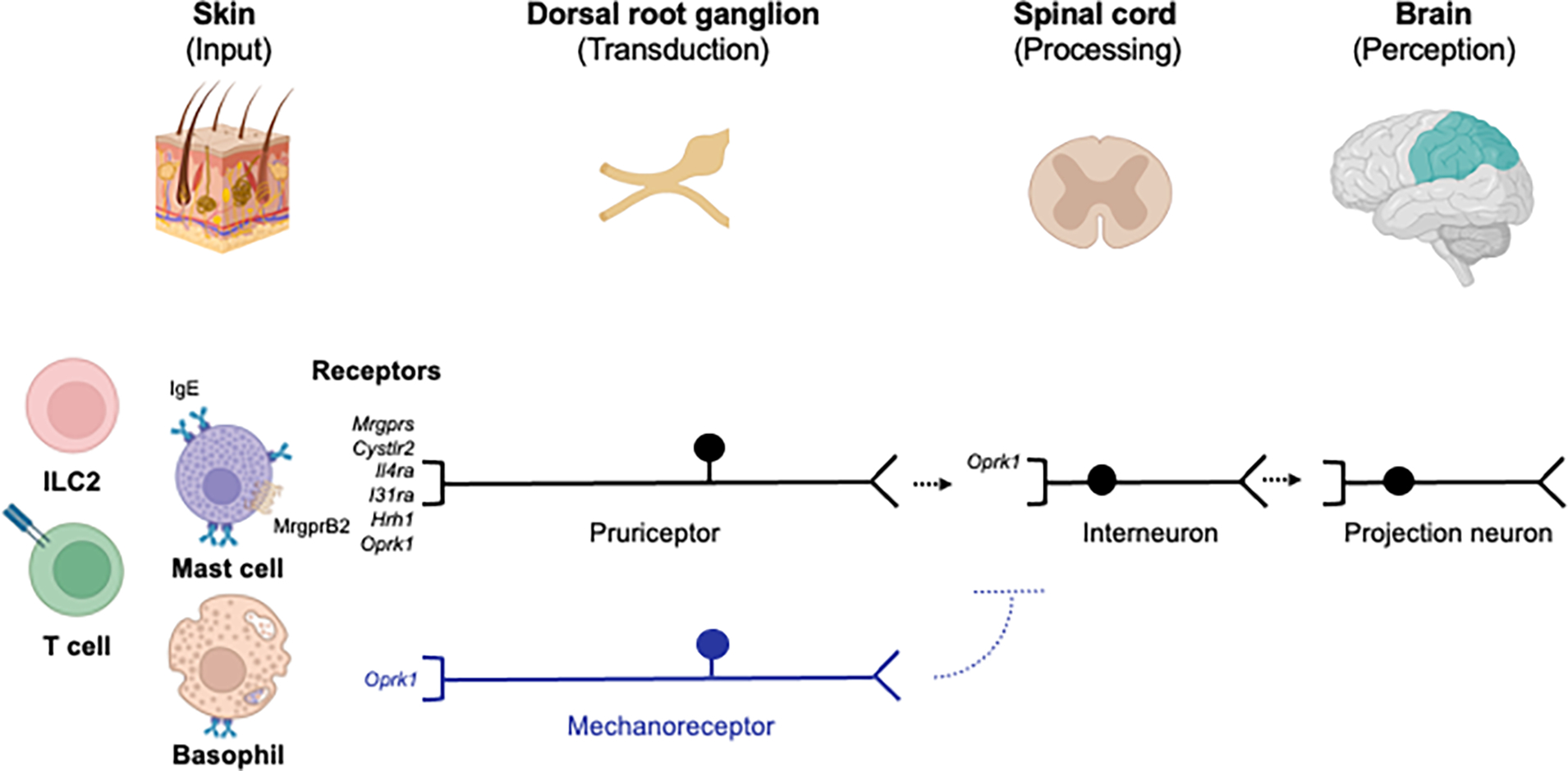
Figure 1. A schematic of basic neuroimmune itch circuitry.

Tissue-resident immune cells in the skin can be activated by a variety of signals including cytokines and/or antigen. Mast cells can be activated by endogenous or exogenous factors via MRGPRX2 and/or IgE and release various pruritogens. These include molecules such as neurotransmitters, neuropeptides, leukotrienes, and cytokines that can activate receptors on pruriceptors encoded by Mrgprs, Cysltr2, Il4ra, Il31ra, or Hrh1 in the skin. These signals stimulate peripheral neuronal transmission to the spinal cord and onto the brain. Other receptors function to suppress itch such as the kappa opioid receptor (KOR) encoded by Oprk1 which is expressed on C fibers, mechanoreceptors, and interneurons. MrgprB2 - Mas-related G protein-coupled receptor (MRGPR) B2; IgE – immunoglobulin E; *Cysltr2 – cysteinyl leukotriene receptor 2; *Il4ra – interleukin 4 receptor; *Il31ra – interleukin 31 receptor A; Hrh1 – histamine H1 receptor; ILC2 – group 2 innate lymphoid cell. *Receptors more specifically expressed on pruriceptors. Created with Biorender.com.
