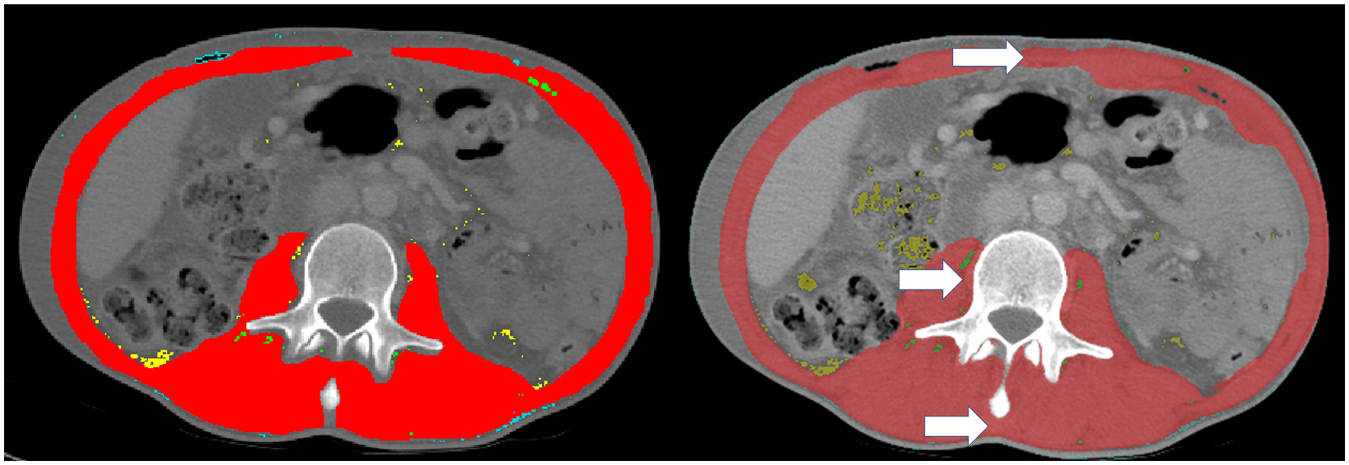
Figure 2:

Representative manual segmentation (A) and automatic segmentation (B) of muscle cross sectional area (red). White arrows depict areas of routinely overestimated muscle tissue at linea alba (top), spinous process (bottom), and tissue beside vertebral body (left). Uncommonly overestimated areas include enhanced intraperitoneal fat (yellow arrow).
