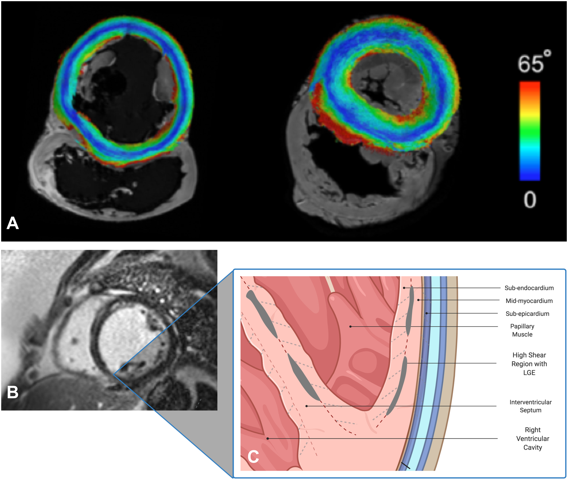
Figure 1:

Panel A: Short-axis slice of normal porcine (left) and human (right) hearts on diffusion-tensor CMR with submillimeter resolution showing various myofiber orientations of different myocardial layers from endo- to epicardium (color-coded by the absolute values of the inclination angle 0–65°). Panels B & C: Short-axis CMR image of a patient with dilated NICM showing linear, mid-wall LGE in the septum, inferior and inferolateral walls. In systole, significant shear forces develop along the myocardial sheets cleavage planes with various orientations (dashed lines). The strain created as these fibers separate leads to increased extracellular space and gadolinium accumulation in the mid-myocardium. This process may be exaggerated in patients with non-ischemic cardiomyopathy.). Created with BioRender.com.
