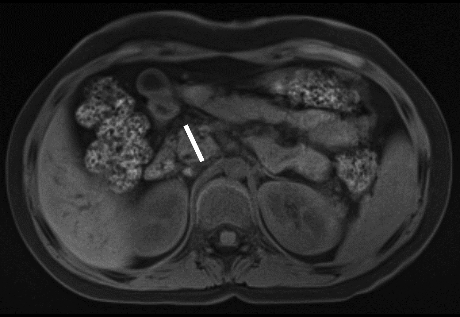
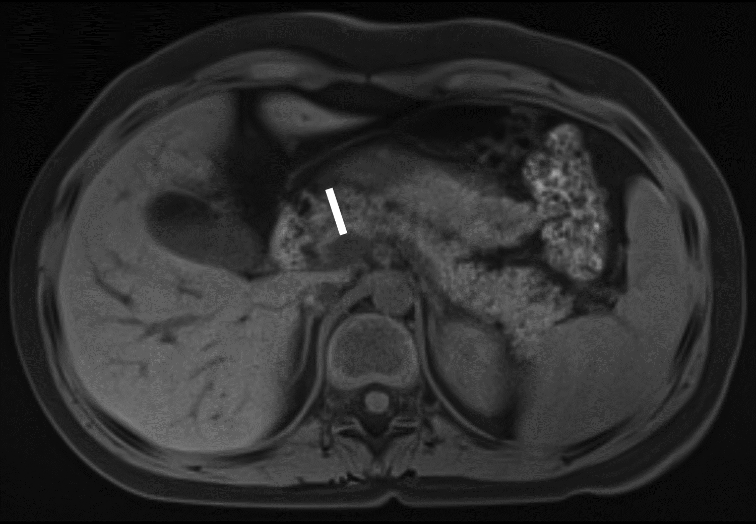
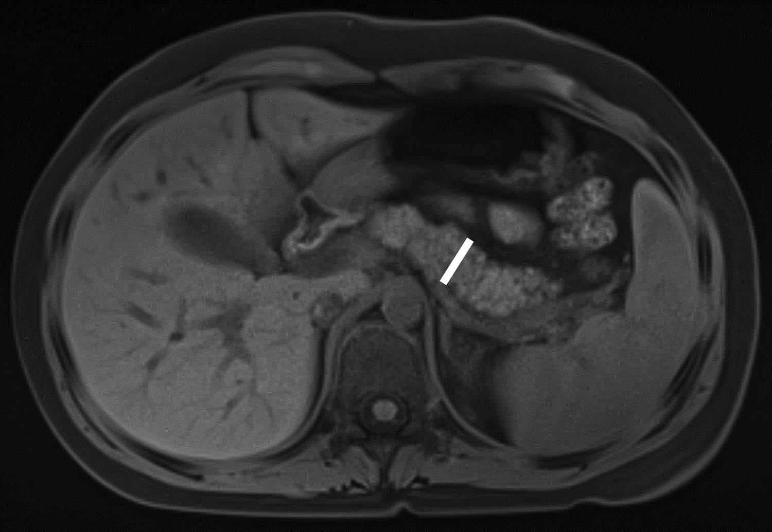
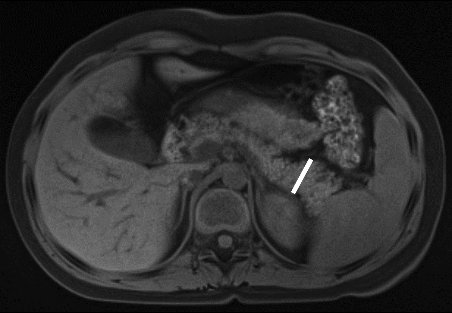
Figure 1 –


Axial T1-weighted fat-saturated gradient recalled echo MR images in 10-year-old girl with chronic pancreatitis, showing measurement of pancreas parenchymal thickness. As previously described [8], measurements were made perpendicular to the surfaces of the pancreas to the right of the superior mesenteric vein and anterior to the inferior vena cava for the head (A), anterior to the superior mesenteric vein for the neck (B), anterior or to the left of the abdominal aorta for the body (C), and 1.5 cm to the right of the tip for the pancreatic tail (D).
