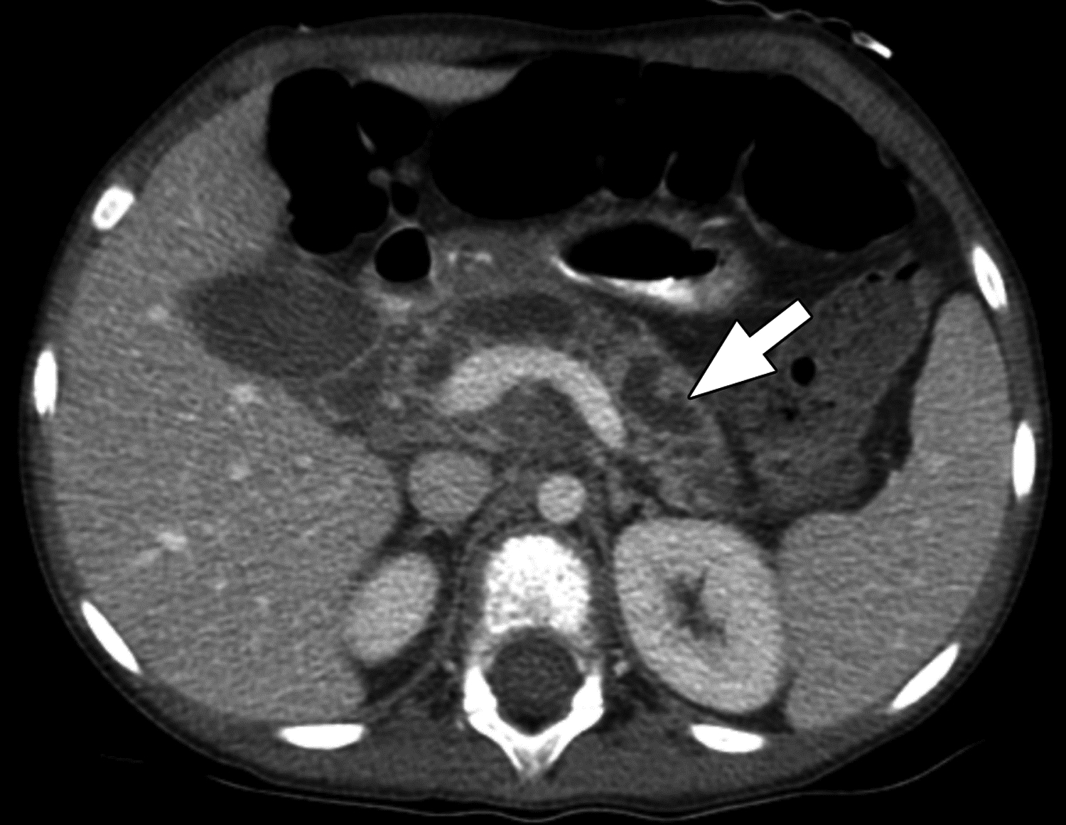
Figure 2 –

Axial contrast-enhanced CT images in 7-year-old girl with chronic pancreatitis. The main pancreatic duct is severely dilated (arrow), and the surrounding pancreatic parenchyma is diffusely thinned (atrophy). The central reviewer and all three site radiologists agreed on the presence of main pancreatic duct dilation and pancreas atrophy.
