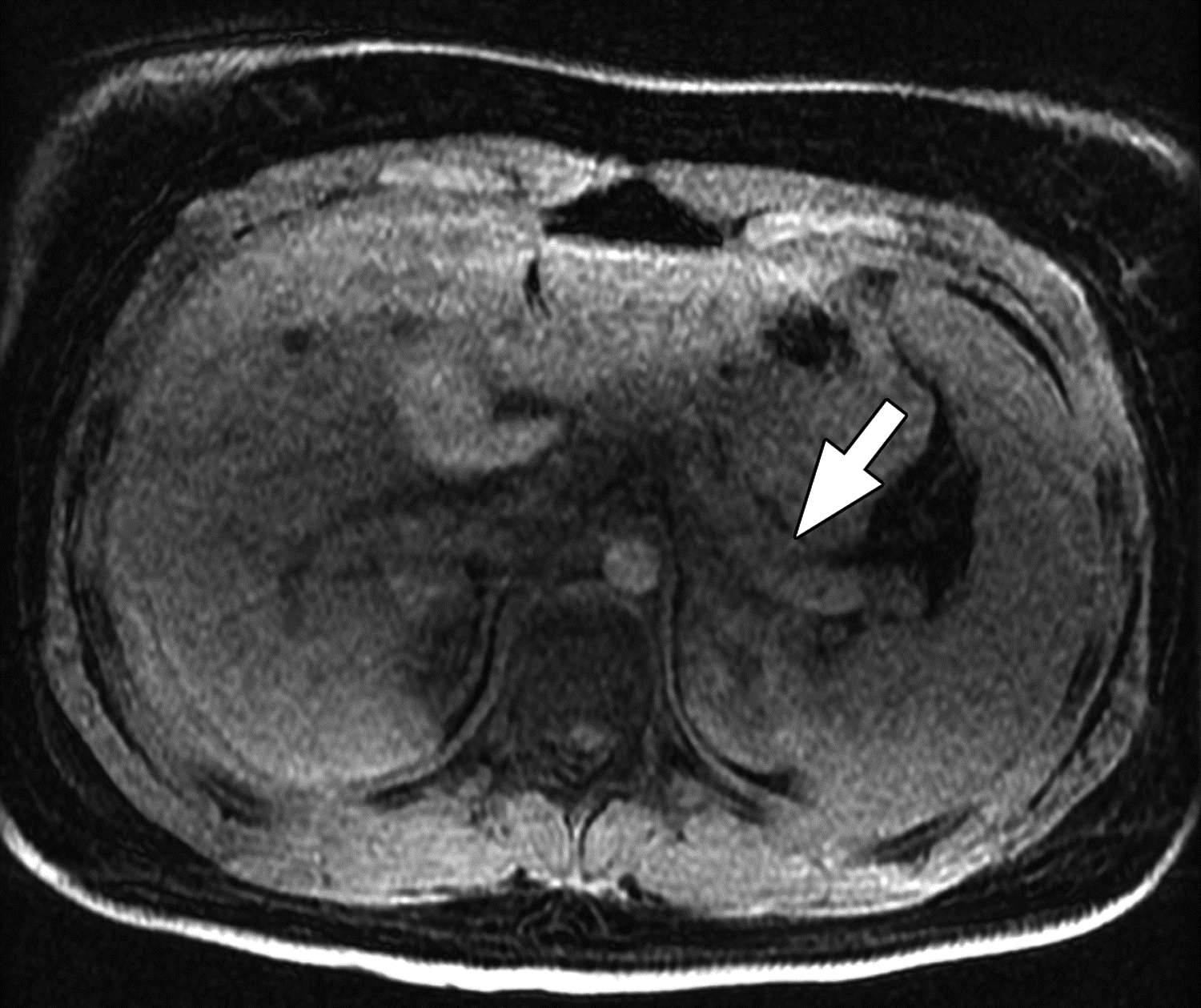
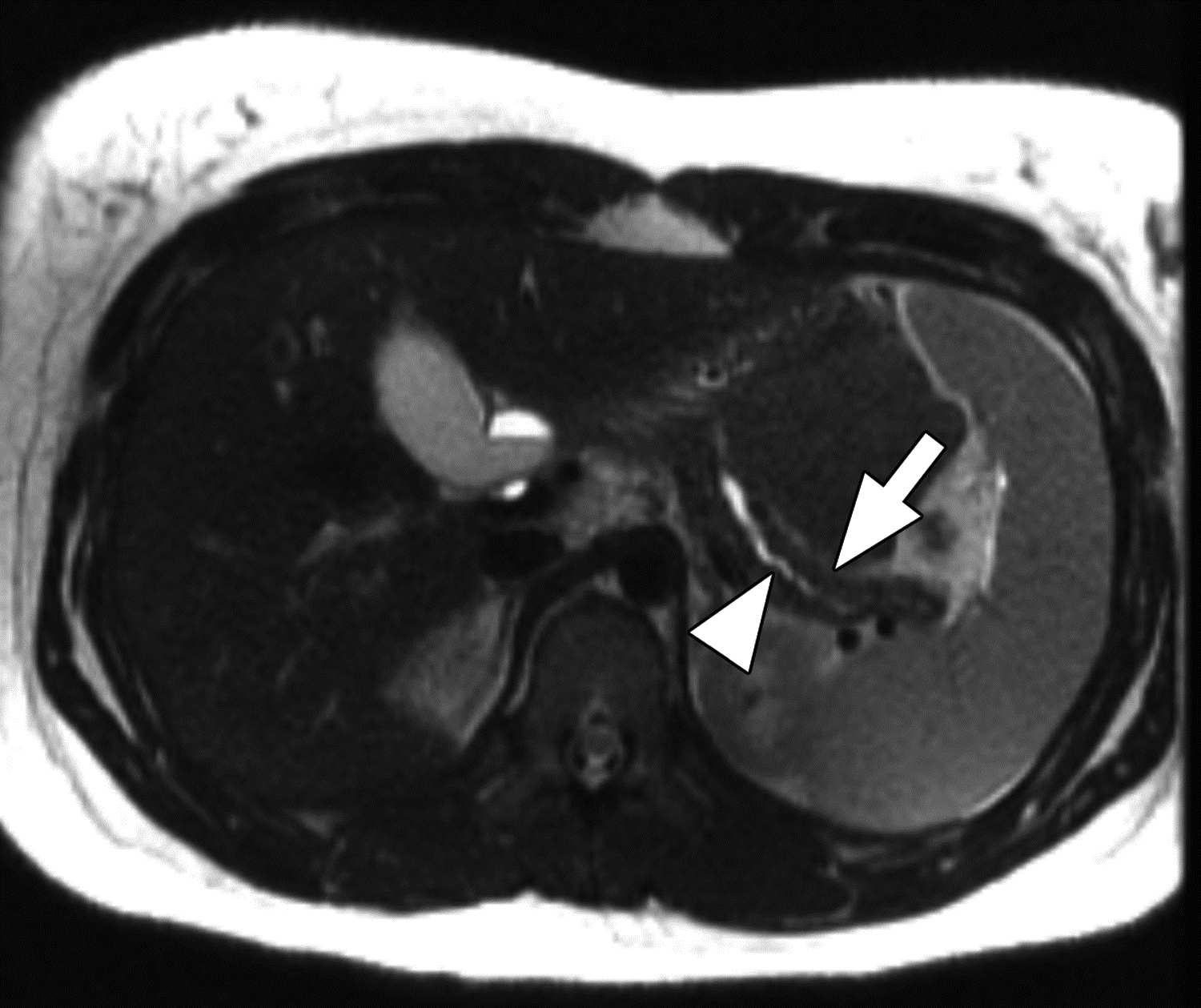
Figure 5 –


MR images from 14-year-old girl with chronic pancreatitis. (A) Axial T1-weighted 3D fast spoiled gradient-echo (FSPGR) water image shows pancreas atrophy (arrow) and heterogeneous loss of T1-weighted signal. (B) Axial T2-weighted SSFSE image shows pancreas atrophy (arrow), main pancreatic duct dilation, and irregularity (arrowhead). The central reviewer and all three site radiologists agreed on the presence of main pancreatic duct dilation, main pancreatic duct irregularity, pancreas atrophy, and loss of T1-weighted signal.
