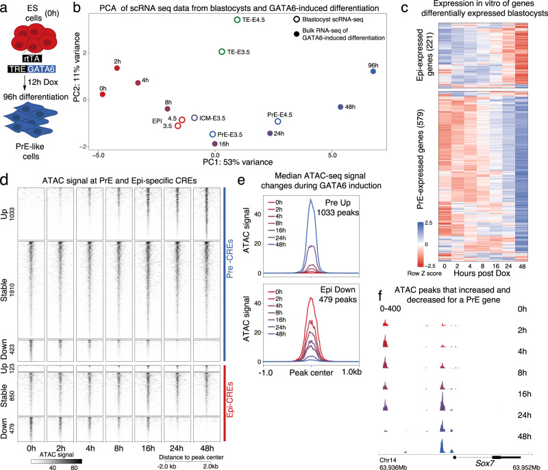
Fig. 1. GATA6-driven in vitro differentiation recapitulates PrE specification in blastocysts and initiates rapid transcriptional changes and chromatin remodeling.
a Scheme depicting GATA6-driven in vitro differentiation of mES cells used to study PrE specification. b PCA of bulk-RNA-seq at differentiation time-points (solid circles) and single-cell RNA-seq profiles of E3.5 and E4.5 blastocysts (open circles). The PrE precursor state was achieved by 16 h of GATA6-driven differentiation and fully specified PrE-cells appeared at 24-48 h. c Heatmap of transcript z-scores of Epi- and PrE-specific genes shows that most blastocyst lineage-specific genes showed similar changes in expression during GATA6-driven differentiation. d Changes in chromatin accessibility measured by ATAC-seq at PrE and Epi CREs were quickly initiated within 2 h of differentiation. Accessible regions at 48 h were compared to 0 h and classified as up, stable or down. e Median ATAC signal plots depict an increase in ATAC signal at 1033 PrE-CREs and decrease at 479 Epi-Cres. f Browser view at the Sox7 locus showing a progressive loss in ATAC signal at a distal CRE and a gain in accessibility at a proximal CRE. This example shows that not all PrE CREs gained accessibility during PrE-specification.

